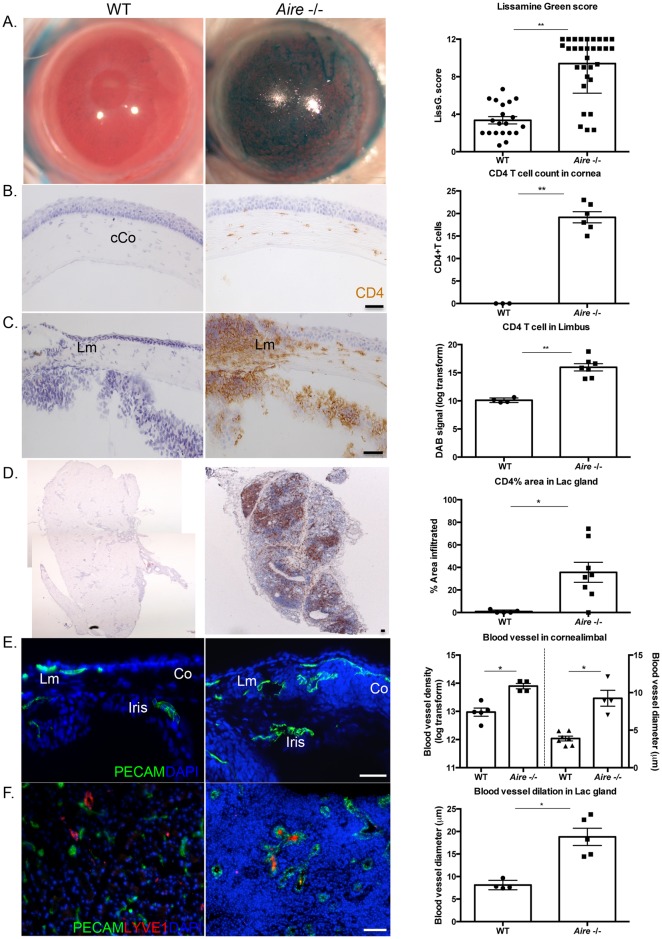
Fig 1. CD4 T cell-mediated inflammation and neovascularization in Aire -/- mice.
(A) Lissamine green staining indicates significant corneal damage in the Aire -/- mice. (B-D) CD4 T cells extensively infiltrate the central cornea (cCo), limbus (Lm), and lacrimal glands of Aire -/-. (E) Blood vessels are increased in number and dilated in the corneolimbal region of Aire -/-. (F) Lymphatic vessels are closely associated with dilated blood vessels in the Aire -/- lacrimal gland. Data are expressed as mean±SEM and are representative of measurements obtained from at least three independent eyes/glands. n ≥ 3 per group. ** p < 0.001, *p < 0.05. Scale bar = 50 μm.

