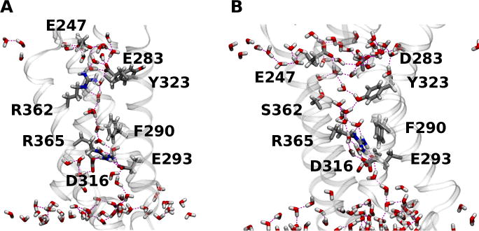
Figure 6.

(A) Configuration snapshot of the native Shaker VSD; and (B) the BOM VSD in the KCl simulation. The side chains involved in interactions with the permeating ions in the BOM mutant VSD systems, the BOM S362 side chain, and the wild-type R362 and E283 side chains are shown as licorice. Waters in the first two solvation shells around the VSD and within 15 Å from the lipid bilayer center are shown in licorice. H bond interactions are drawn as broken lines.
