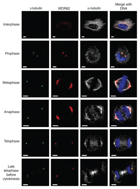
Figure 2.
Subcellular localization of WDR62 throughout the cell cycle. Confocal microscopy analysis of HeLa cells during each stage of the cell cycle. WDR62 staining is weak and cytoplasmic during interphase, with a concentration at a perinuclear position suggestive of the Golgi apparatus, and shows spindle pole localization during mitosis. Cells were stained with antibodies against human WDR62 (red), γ-tubulin (green) as a centrosome marker, α-tubulin (white) as a microtubule marker, and DNA (blue) stained with DAPI. Scale bar, 5 μm.

