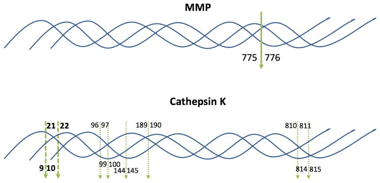
Fig. 1.
Schematic representation of MMP and cathepsin K cleavage sites in type I collagen. The bold, solid green arrow indicates the known MMP cleavage site (bond 775–776) aligned in all three chains in the triple-helix. The bold, dashed green arrows indicate the cathepsin K cleavage sites where all three chains in the triple-helix align (bonds 9–10 and 21–22; see Table 1). The dashed green arrows indicate the cathepsin K cleavage sites in individual collagen chains that do not align within the triple-helix (see Table 1). For cleavage by cathepsin K within individual chains, sites in the α1(I) chain are noted above the triple-helix, while cleavage sites in the α2(I) chain are noted below the triple-helix.

