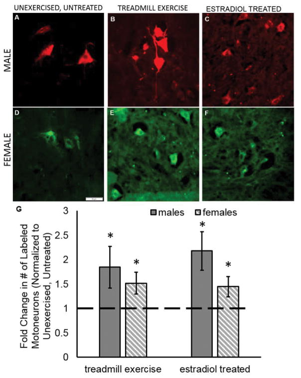
Figure 3.
Exercise and estradiol treatments enhance the number of motoneurons that participate in regeneration in males and females. Sample pictographs from unexercised, untreated male (A) and female (D), treadmill exercise treated male (B) and female (E), and estradiol treated male (C) and female (F). Motoneurons were labeled 1.5 mm distal to the original cut site using either AlexaFluor 594 (red) or AlexaFluor 488 (green) conjugated tracers. Both retrograde tracers were used in both sexes, and no differences were found in the number of cells counted within a treatment group when separated by tracer. Scale bar 50 μm. (G) Treatment with estradiol or exercise using either the continuous paradigm (males; sold bar) or interval paradigm (females; hashed bar) significantly increased the number of labeled motoneurons compared to unexercised, untreated controls (dashed line). Data are normalized to unexercised, untreated control motoneuron counts. Each sex was normalized to the controls of the same sex. Errors bars, ± standard deviations. * p ≤ 0.001

