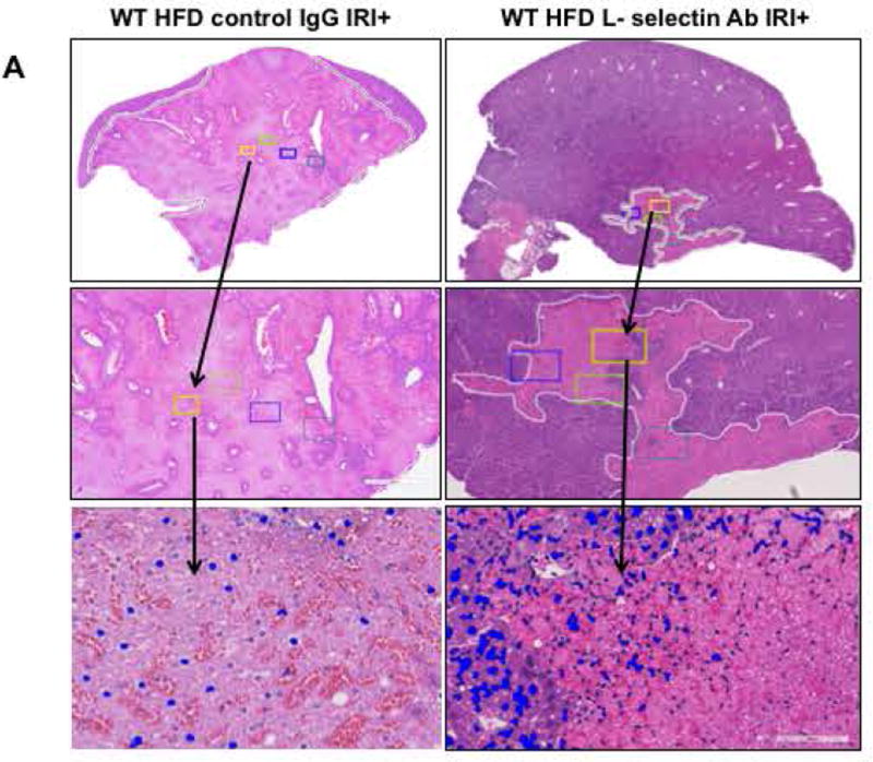
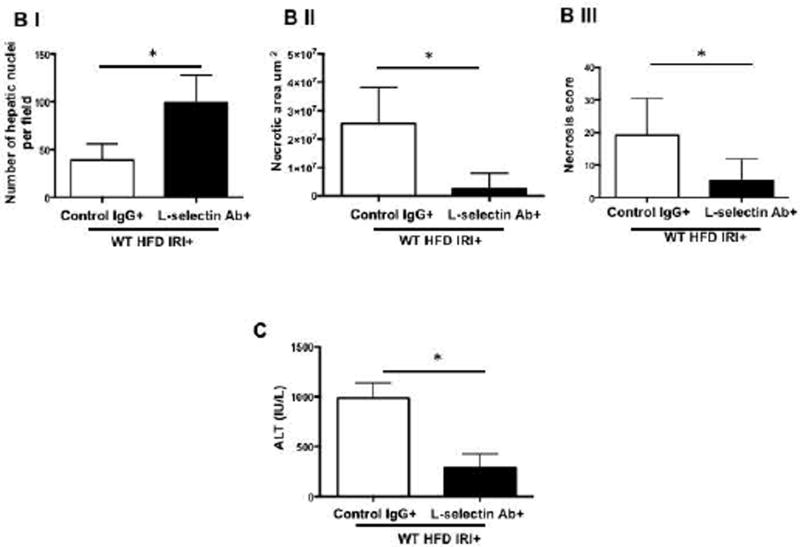
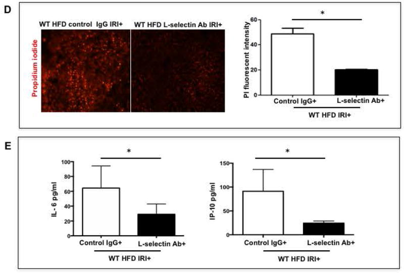
FIG. 5. Blocking L-selectin reduces hepatocellular injury in HFD fed mice after IRI.
(A) Histological representation of ischemic liver lobes obtained from WT HFD fed mice treated with control IgG and L-selectin antibody and subjected to IRI. Entire lobe of the liver undergoing IRI was scanned and representative H&E images demonstrating area of necrosis (outlined by white lines) are shown here. Middle panels represent magnified images of top panels and bottom panels represent hepatic nuclei (blue). (BI,II) Control IgG vs. L-selectin Ab for hepatic nuclei / field (p<0.0001) and necrotic area (p<0.005) (BIII) Necrosis score (p<0.002) (C) ALT (p<0.004) (D) depicts extent of necrotic cell death by red fluorescent stain, Propidium iodide (PI) positive cells in the liver from HFD IRI+ given control IgG and L-selectin Ab .PI intensity quantification shown on right (p<0.003). (E) Quantification of serum IL-6 (p<0.04) and IP-10 (p<0.03) from IgG and L-selectin Ab treated HFD fed mice. Data represents mean ± SEM of at least 10 mice in each group of 3 independent experiments. Asterisks indicates p<0.05, student t-test.


