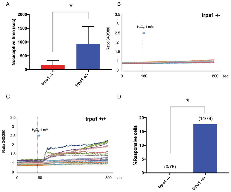
Fig. 7. H2O2-induced nociceptive behavior and Ca2+ transients in dorsal root ganglia (DRG) neurons induced by H2O2 in TRPA1 knockout (TRPA1 −/−) and wild-type (TRPA1 +/+) mice.
(A) Total time of nociceptive behavior over 60 minutes after H2O2 (0.05 ml, 30 mM) was injected into the gastrocnemius muscle. Data were obtained from eight animals in each group. * P = 0.0051 compared with the trpa1 +/+ group by unpaired t-test. Data are expressed as means ± SD. (B) Example traces in response to H2O2 (1 mM; 20-second duration) of individual, dissociated lumbar 3–5 DRG neurons from TRPA1 −/− mice during Fura-2 Ca2+ imaging. (C) Example traces in response to H2O2 (1 mM; 20-second duration) of individual, dissociated lumbar 3–5 DRG neurons from TRPA1 +/+ mice during Fura-2 Ca2+ imaging. (D) Summary of the percentage of cells responding to 1 mM H2O2. Numbers in parentheses represent the number of cells responding and total number of cells tested. * P < 0.0001 compared with the TRPA1 +/+ mice group by Chi-square test.

