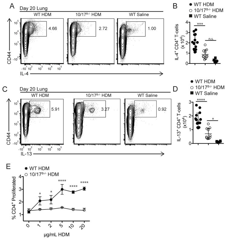
Fig. 6. ADAM10/17 B−/− mice have decreased TH2 responses in a HDM allergic airway hypersensitivity model.
(A–D) At day 20, lungs were digested as described in methods and stimulated with plate-bound anti-CD3 (2μg/mL) for 4 hours in the presence of monensin. Relative (A, C) and absolute (B, D) numbers of IL4+ (A, B) and IL13+ (C, D) effector T cells were determined by intracellular staining and flow cytometry. (E) medLN cells were isolated at day 20 and restimulated with increasing concentrations of HDM for 3 days. Proliferation was measured by cell trace violet. n.s., not significant (P ≥ 0.05). *P < 0.05, **P < 0.01, ***P < 0.001, ****P < 0.0001. One-way analysis of variance (ANOVA) with Tukey’s post-test (B, D), two-way ANOVA with Tukey’s post-test (E). Data are pooled from three (A–E) independent experiments.

