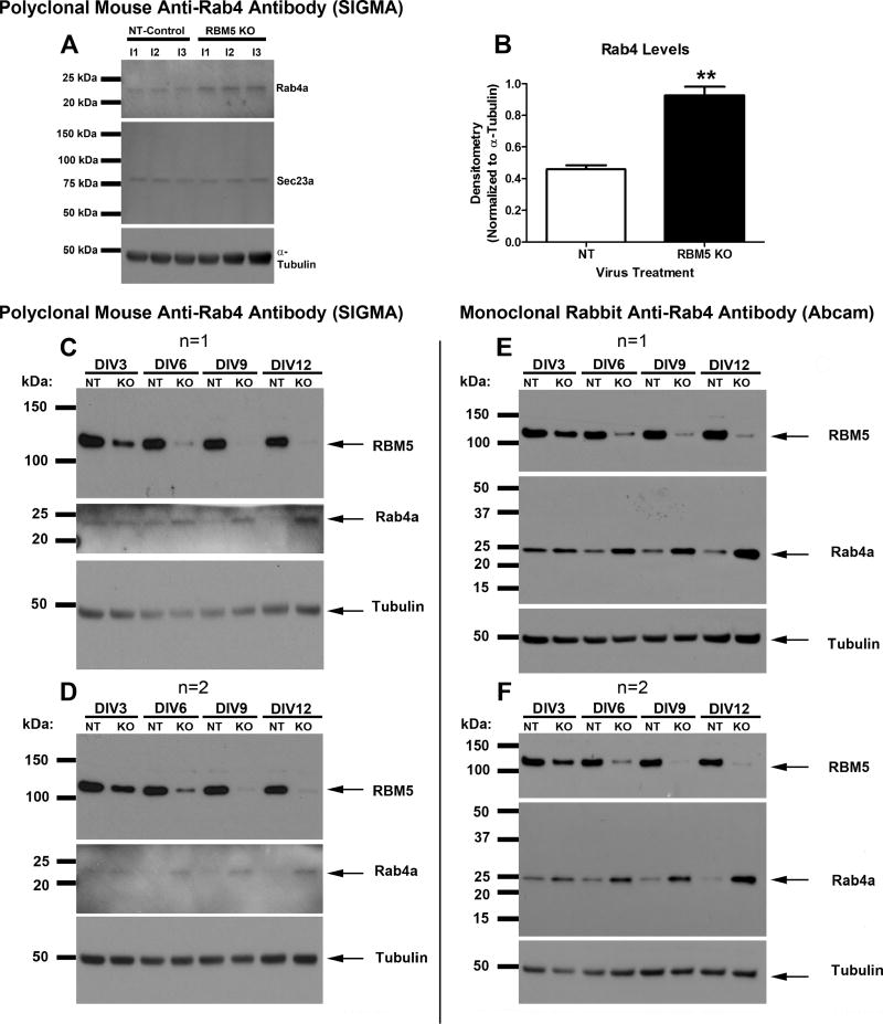
Figure 4. Rab4a Protein is increased in KO vs. Control Samples as Measured by Two Different Antibodies.
(A) Western blots show that Rab4a protein (but not Sec23a) is increased in KO neurons relative to controls in three independent neuron culture isolation experiments (i1, i2, i3), as detected by a mouse polyclonal antibody (SIGMA). (B) Densitometry shows significant increase in Rab4a levels in KO neurons vs. controls. (C–F) Neurons were transduced with lentivirus to overexpress NT-shRNA or RBM5 targeting shRNA and protein extracts were harvested between DIV3-DIV12. Two biological replicates (n=1 and n=2) were prepared/harvested from a single neuron culture isolation experiment. (C and D) Age course samples were probed with anti-RBM5, anti-Rab4 (SIGMA, Mouse Polyclonal), and anti-α-Tubulin antibodies. (E and F) Age course samples were probed with anti-RBM5, anti-Rab4 (Abcam, Rabbit Monoclonal), and anti-α-Tubulin antibodies. Data were analyzed by unpaired T-test. Data were significant at p <.05 (*). Graphs show mean + SEM.

