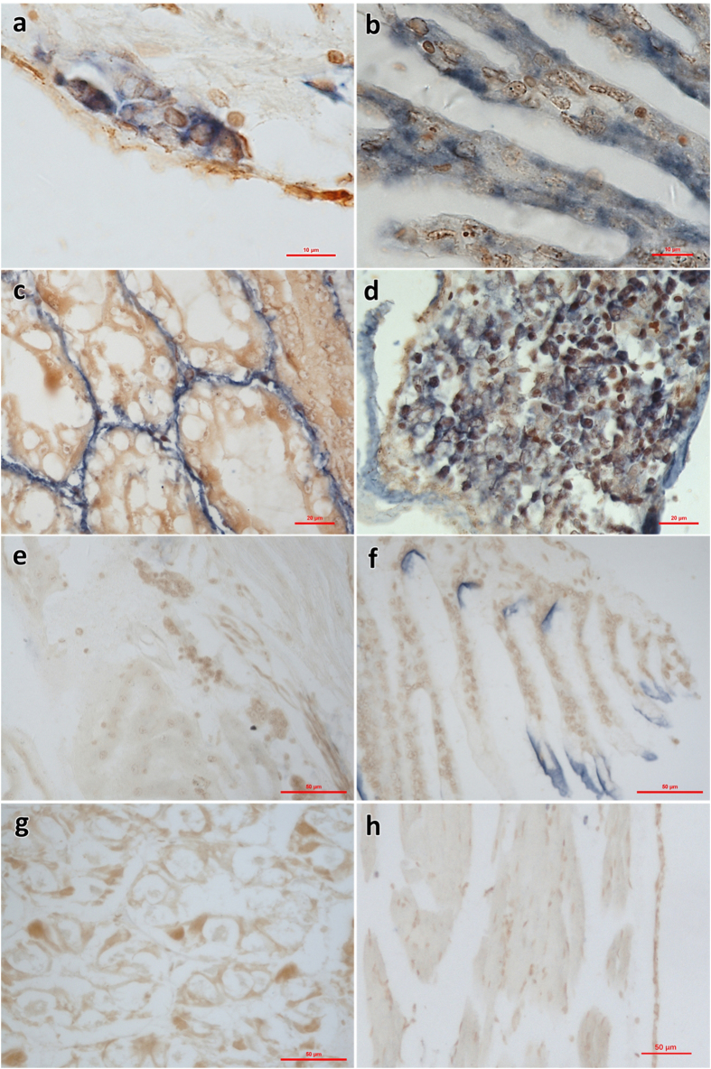
Figure 7.
ISH using a digoxigenin labeled 279 bp probe for the shrimp hemocyte iridescent virus on histological sections of L. vannamei. (a)–(d) hematopoietic tissue, gills, hepatopancreas and periopods in SHIV-positive sample, respectively; (e)–(h) hematopoietic tissue, gills, hepatopancreas and periopods in SHIV-negative sample, respectively. In (a)–(d), blue signals were observed in the cytoplasm of the hemocytes of hematopoietic tissue, gills, sinus of hepatopancreas and periopods. In (e)–(h), no hybridization signal was seen on the same tissues of SHIV-negative L. vannamei except some non-specific signals on cuticle. Bar, 10 μm (a and b), 20 μm (c and d), and 50 μm (e–h), respectively.

