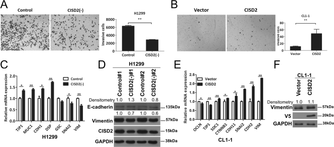
Figure 4.
CISD2 promotes cell invasion in lung ADC cells. (A and B) Left panel, representative images of cell invasion assay showing invading cells on the bottom side of membrane of transwells; right panel, result of quantification analysis. (C) RT-qPCR analyses of the transcript levels of EMT markers in shCISD2 transfectant cells. (D) Western blot analysis of EMT marker proteins in shCISD2 transfectant cells. (E) RT-qPCR analyses of the transcript levels of EMT markers in CISD2(+)-CL1-1 cell. (F) Western blot analysis of EMT markers in CISD2(+)-CL1-1 cells. For (A), (B), (C) and (E), p values were obtained using Student’s t-test, *P < 0.05, **P < 0.01. All data are mean ± SD of at least triplicate measurements.

