Figure 3.
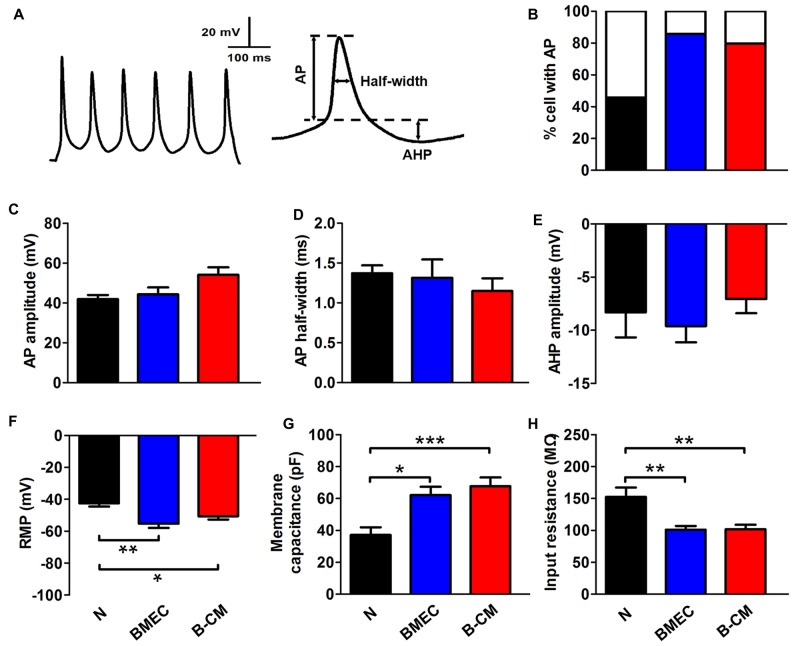
BMEC promote electrophysiological development in cortical neurons. (A) The typical whole-cell current clamp recordings with depolarizing current (50 pA) injection in cortical neurons co-cultured with BMEC for 2 days. Scale bar is shown in the top. The right panel represents action potential (AP) measures taken. (B) Summarized data show that significantly more neurons display AP spikes when grown with BMEC or treated with B-CM than those cultured alone (N) (N: n = 24; BMEC: n = 21; B-CM: n = 24. N vs. BMEC, p < 0.05; N vs. B-CM, p < 0.05). (C–E) The panels represent the measurements taken for AP amplitude, AP half-width and after hyperpolarization (AHP) amplitude respectively. There is no significant difference of AP properties among three groups (N: n = 9; BMEC: n = 16; B-CM: n = 14). (F–H) Summary graphs showing the passive membrane properties (Resting membrane potential (RMP), membrane capacitance (Cm) and input resistance (Rin)) of cortical neurons grown alone, co-cultured with BMEC or treated with B-CM (n = 10 per condition. *p < 0.05; **p < 0.01; ***p < 0.001).

