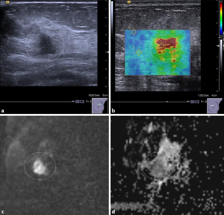
Fig. 1.
A 48-year-old woman with invasive ductal breast cancer, G3. The lesion (dashed circle) presents as an ill-defined hypoechogenic lesion on B‑mode ultrasound (a) that is associated with high SWV (4.6 m/s), coded red on the parametric ARFI overlay (b). The MRI-DWI scan of the same lesion shows a hyperintense lesion (c) corresponding to restricted diffusivity (1 * 10−3 mm2/s) that appears dark on the quantitative ADC map (d)

