Fig. 6.
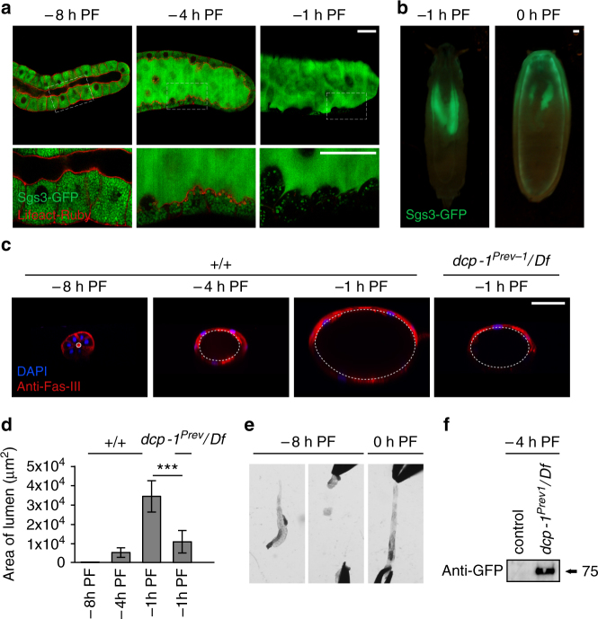
Cortical caspase activation in living salivary glands controls tissue elasticity during expulsion of mucin-like glue proteins. a Dismantling of cortical F-actin in acinar cells coincides with secretion of glue proteins; boxed areas imaged at higher magnification below. At −8 h PF, glue proteins (Sgs3-GFP, in green) are present only in the cells of the salivary gland. At −4 h PF, exocytosis of glue proteins from cells into the lumen begins; by −1 h PF, all glue is present in the lumen. Lifeact-Ruby, in red, shows that F-actin begins to break down at −4 h PF, and dismantling is nearly complete by −1 h PF. Salivary glands were imaged live/unfixed. b Glue proteins present in the lumen at −1 h PF are expelled onto the surface of the animal at 0 h PF. c, d The lumen increases dramatically in size during glue exocytosis, and luminal expansion requires caspase activity. c Transverse view of salivary glands shows luminal expansion during glue secretion. Fasciclin-3 staining (anti-Fas-III, in red) marks septate junctions, nuclei stained with DAPI in blue. At −8 h PF, salivary glands have a narrow lumen; however, as glue exocytosis progresses, the lumen expands dramatically until reaching maximal size at −1 h PF. The lumen of dcp-1 mutant salivary glands does not expand normally. d Quantification of luminal area for stages and genotypes shown in c (n = 10 per timepoint, error bars indicate s.d., asterisks indicate p < 0.01 determined by one-tailed t-test). e Salivary gland elasticity is developmentally controlled. At −8 h PF, salivary glands are rigid and tear at the slightest pull. In contrast, 0 h PF salivary glands can be stretched beyond their normal length. Equal force was applied to each stage; n = 20 tested per stage. f Luminal expansion is critical for timely expulsion of glue proteins. Glue expulsion normally occurs after larvae become stationary. Control wandering larvae at −4 h PF do not expel glue, while dcp-1 mutant larvae precociously expel glue at this stage. Expelled glue proteins (in Sgs3-GFP animals) were collected and measured by western blot with anti-GFP antibodies. Scale bars represent 100 µm. Df, deficiency, PF, puparium formation

