Figure 5.
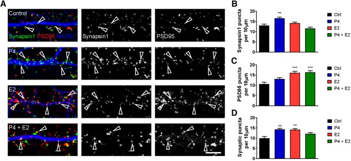
Progesterone (P4) and estradiol (E2) alter synapse number and synaptic protein expression. A, Representative confocal images of primary rat cortical neurons treated with 1 nm P4, 1 nm E2, or both for 24 h. Images are for synapsin-I (green) and PSD95 (red) along MAP2-positive dendrites (blue). Open arrowheads indicate colocalized synapsin-I and PSD95 puncta and thus, synapses. B, Quantitative analysis of synapsin-I linear density; treatment with P4 increased synapsin-I density compared with control (vehicle) condition. C, PSD95 linear density was increased over control levels after treatment with E2 alone or combined P4 and E2 treatment. D, Measurement of synaptic puncta, defined as synapsin-I puncta positive for PSD95, was increased after treatment with either P4 or E2. **p < 0.01; ***p < 0.001; scale bar = 5 µm.

