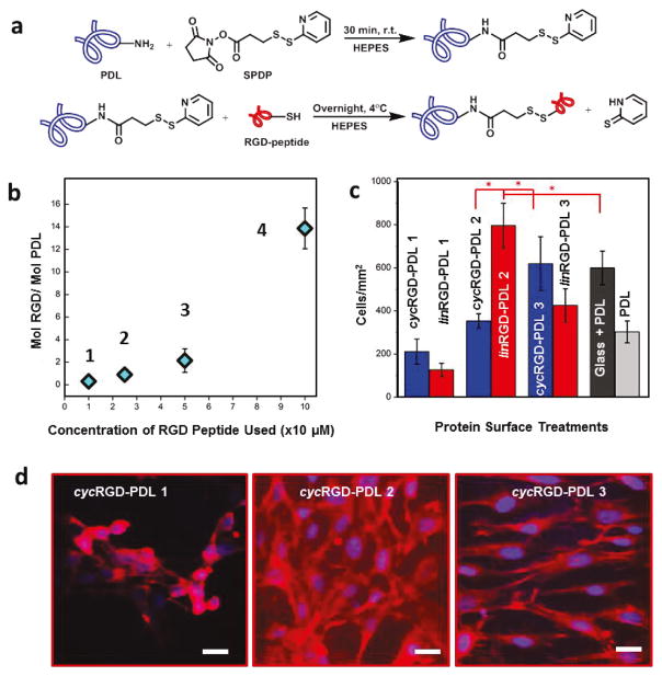
Figure 1. Modified PDL Synthesis and Assessment of RGD-dependence of Cell Growth and Morphology using NIH/3T3 Embryonic Murine Fibroblasts.
(a) Covalent coupling mechanism between PDL and RGD-containing peptides. (b) Plot illustrating cyc(lin)RGD-PDL 1–4 as the degree of the RGD modification of PDL. Error bars represent standard deviations and are smaller than data point diamonds for 1 and 2. (c) NIH/3T3 fibroblast cell densities at 24 hours in culture on different surface treatments. Tukey’s means comparisons (Supplementary Information, Table S1) are highlighted by red asterisks, p < 0.01. (d) CFM Images of 3T3 fibroblasts on representative surface treatments 24 hours after seeding. Actin filaments stained with rhodamine-phalloidin (red), and nuclei stained withDAPI (blue). Scale bar is 15 μm.

