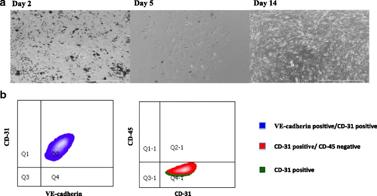
Fig. 1.

Isolation and Culture of Primary Ovarian Endothelial Cells. a. Mouse ovarian ECs (CD31+) were isolated using magnetic cell sorting and cultured in serum free media. 7 days later, activation of the ECs was initiated with lentivirus transduction of the myr-Akt gene. 14 days post isolation, AOEC are proliferating. b. FACS analysis of AOEC isolated by magnetic cell sorting, cultured on serum free media, passage 6. 98% of the cells were CD 31 positive, VE-Cadherin positive and CD45 negative, matching a known EC profile. This experiment was repeated 5 times; all cultures contained at least 93% fully matched endothelial cells
