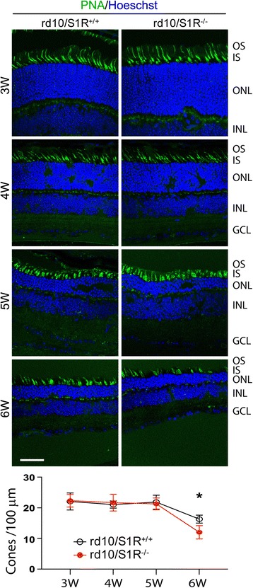
Fig. 1.

PNA label indicating exacerbated cone loss due to S1R knockout in rd10 mice. Animals were euthanized at indicated time points, and retinal sections were prepared for cone labeling with fluorescent PNA. OS/IS: outer/inner segment; ONL: outer nuclear layer; INL: inner nuclear layer; GCL: ganglion cell layer. Scale bar: 20 μm. Quantification: cone cell number per 100 μm ONL length; mean ± SE, n = 3–4 mice; *P < 0.05
