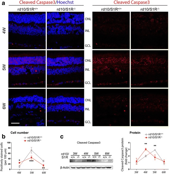
Fig. 3.

Accelerated production of cleaved Caspase3 in rd10 retinas due to S1R knockout. a. TSA-enhanced immunostaining of cleaved Caspase3 on retinal sections collected at indicated time points. The one-color (channel) version is shown on the right. ONL: outer nuclear layer; INL: inner nuclear layer; GCL: ganglion cell layer. Scale bar: 20 μm. b. Quantification of cells stained positively for cleaved Caspase3. Mean ± SE, n = 3–4 mice; *P < 0.05, **P < 0.01, ***P < 0.001. c. Quantification of Western blots showing cleaved Caspase3 in retinal homogenates. For the method of quantification refer to Fig. 2b; mean ± SE; n = 4; **P < 0.01
