Fig. 3.
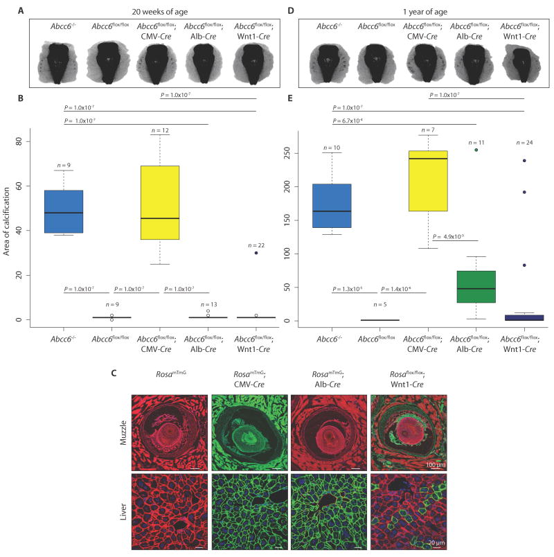
Liver-specific deletion of Abcc6 does not phenocopy constitutive ablation of Abcc6 in mice.
Micro-CT scans of the muzzle to evaluate the extent of vibrissae fibrous capsule calcification were obtained at 20 weeks (A) and 1 year (D) of age. (B and E) Quantification of ectopic calcification from micro-CT images. A one-way ANOVA with Tukey’s honest significance difference post hoc analysis was performed. One-way ANOVA: (B) P = 2.2 × 10⁻¹⁶; (E) P = 4.68 × 10⁻¹¹. P values of post hoc comparisons are indicated in the figure. (C) Visualization and confirmation of Cre-targeted tissues and cell types using the RosamTmG reporter mouse line. All cells that are successfully recombined transition from expression of membrane Tomato (mT; red fluorescence) to green fluorescent protein (mG; green fluorescence). Representative images are shown.

