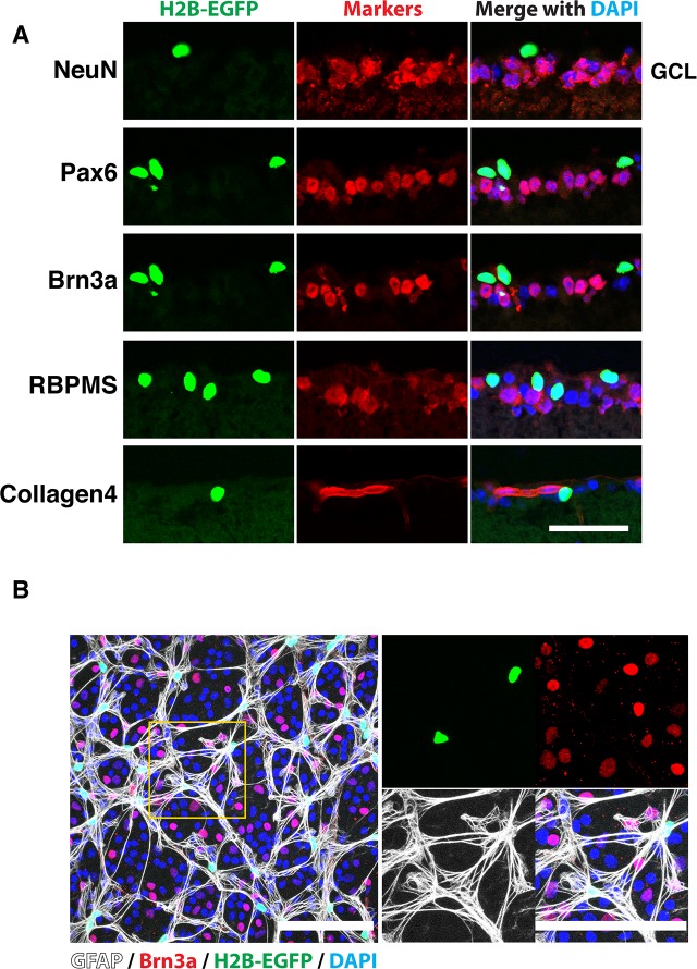
Figure 4.
Colocalization of EGFP fluorescence with a glial marker in the GCL. (A) Transverse sections of 1-month-old αGFP eye were stained with indicated antibodies and DAPI. Only areas corresponding to the GCL are shown. Note the absence of colocalization of EGFP fluorescence with neuronal and blood vessel markers. (B) Whole-mount retina from 1-month-old αGFP/+ mouse was stained with indicated antibodies and DAPI. Right images are enlarged images of the boxed area on the left. Scale bars: 50 μm (A); 100 μm (B).

