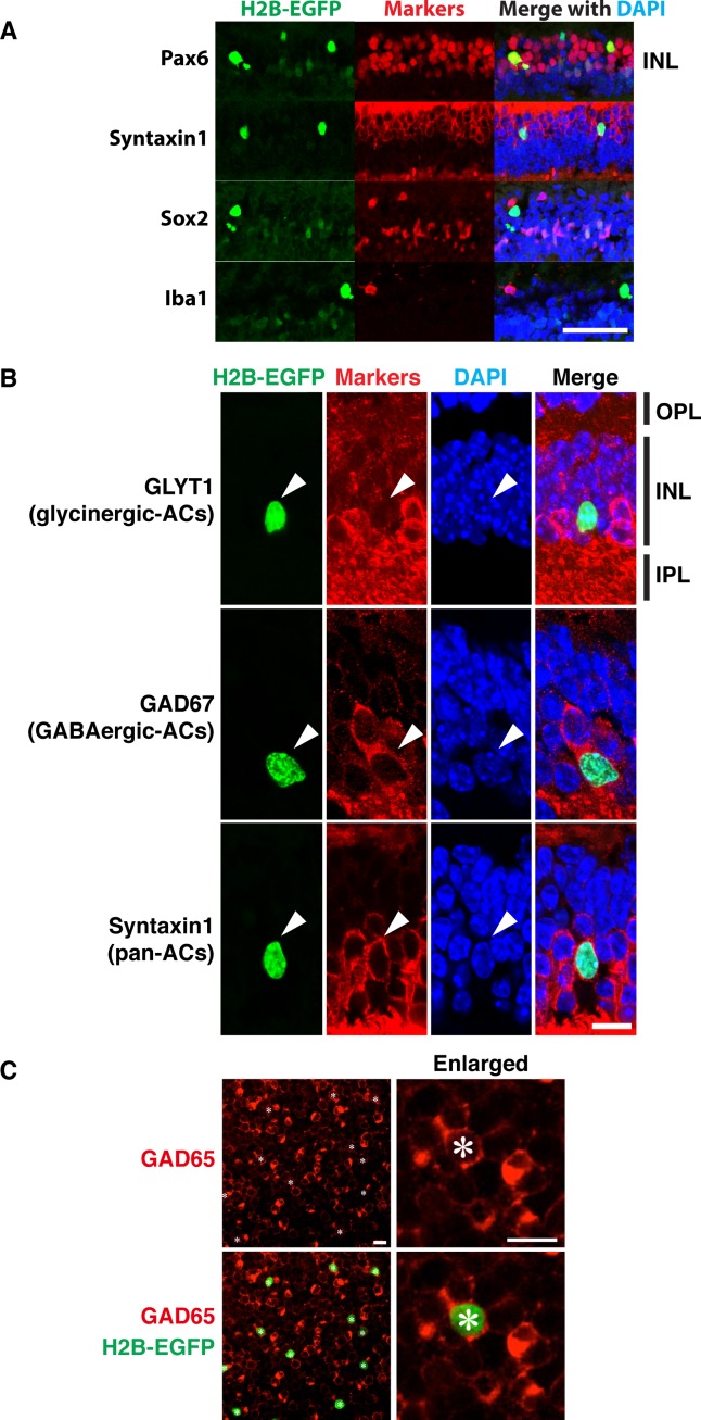
Figure 5.
Colocalization of EGFP fluorescence with amacrine cell markers in the INL. (A, B) Transverse sections of 1-month-old αGFP eye were stained with indicated antibodies and DAPI. Arrowheads mark the position of GFP-positive cells. IPL, inner plexiform layer; OPL, outer plexiform layer. (C) Whole-mount retina from αGFP/+ mouse was stained with antibodies against GAD67 and DAPI. Only GAD67 staining is shown on the right. Asterisk marks EGFP-positive cells. Scale bars: 50 μm (A); 10 μm (B, C).

