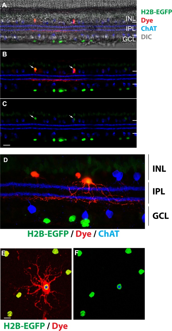
Figure 6.
Identification of amacrine cell type in the inner nuclear layer. (A–C) In a retinal slice, neurobiotin-injected amacrine cells (red) are EGFP positive (green). Starburst amacrine cells are labeled with a ChAT antibody (blue). Arrows point to the soma of amacrine cells. (D) A high-magnification image shows that the ramification of the amacrine cell dendrites (red) is beneath the ON ChAT band (blue). EGFP is shown in green. (E, F) In a piece of whole-mount retina, neurobiotin (red) injected into a EGFP-positive amacrine cell (asterisk) diffuses to neighboring EGFP-positive amacrine cells. Scale bars: 20 μm (A–C); 10 μm (D–F).

