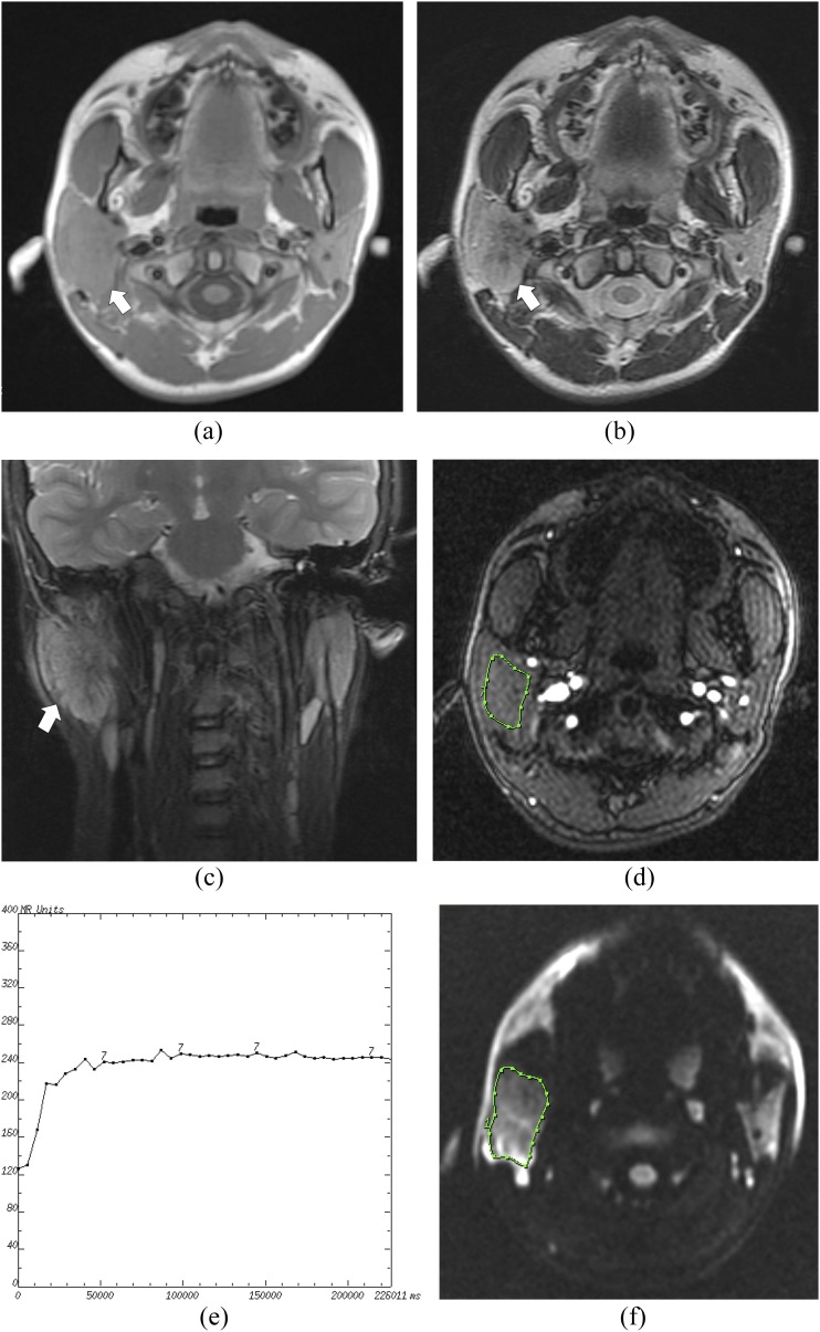
Figure 3.
A mucoepidermoid carcinoma of the right parotid gland in a 15-year-old male: (a) the axial T1 weighted MR image shows an isointense signal mass (arrow) in the right parotid gland with an irregular shape. (b) The axial T2 weighted MR image shows a heterogeneously hyperintense mass (arrow). (c) The coronal T2 weighted MR image shows no capsule around the mass (arrow). (d) The cursor marks the region of interest (ROI) selected for signal intensity (SI) measurement with dynamic MRI. (e) The time–intensity curve shows a plateau enhancement pattern (Type C). (f) The diffusion-weighted image shows a relatively high SI mass. The cursor marks the ROI selected for measurement of the apparent diffusion coefficient (ADC) value. The mean ADC value of this tumour is calculated as 1.01 × 10⁻³ mm2 s−1.

