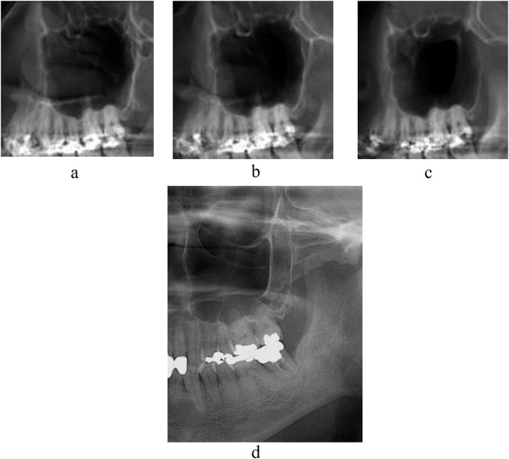
Figure 5.
The inner (a), centre (b) and outer (c) images simulated at the layers of Figure 2 show no evidence of the diagonal line in Case A without maxillary sinus anterior wall depression. On the actual panoramic image (d), the line also cannot be observed.

