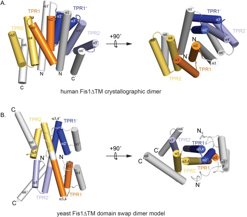
Figure 1.

Structural models for Fis1 dimerization. (A) A cartoon representation shows the dimer model of human Fis1 that requires reciprocating interactions between helix 1 and the TPR concave surface. TPR1 is orange/blue, TPR2 is yellow/light blue and the N‐terminal helices 1 and C‐terminal helices 6 are colored white. The transmembrane domain is not shown. PDB code: 1NZN.pdb.19 (B) A cartoon representation shows the 3D domain swap dimer model of yeast Fis1 cytoplasmic domain which requires a loop to helix transition between TPR1 and TPR2.20 The N‐terminal arm (white) binds into the concave surface, which is comprised of 2 TPR motifs from two protomers (TPR1 orange/blue; TPR2 yellow/light blue). This model was docked using ClusPro.21, 22, 23, 24, 25
