Figure 2.
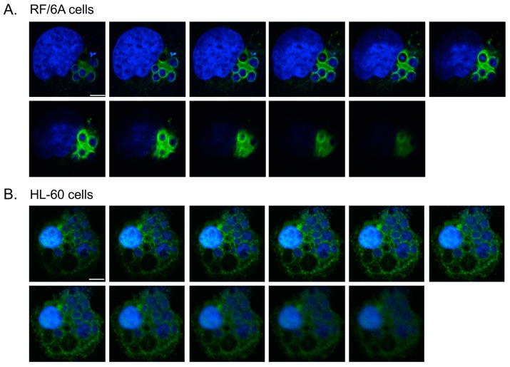
Z-stack analyses confirm vimentin assembly around the ApV. A. phagocytophilum infected RF/6A (A) and HL60 (B) cells were labeled with vimentin antibody and stained with DAPI. LSCM and Z-stack analyses were performed. Scale bars, 5 μm. Results in panels are representative of three experiments with similar results.

