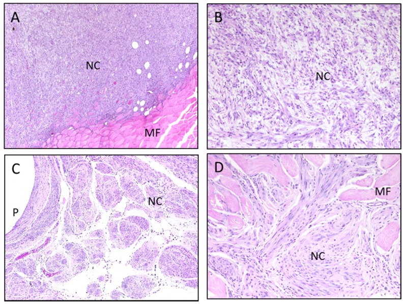
Figure 3.
Histopathologic examination of leg tumors in 24-month metal-implanted mice. (A,B) H&E-stained section of tumor surrounding WTa pellet; (C,D) H&E-stained section of tumor surrounding CoTa pellet; (A,C) magnification ×40; (B,D) magnification ×100. NC, neoplastic cells; MF, normal muscle fiber; P, pellet hole; H&E, hematoxylin and eosin; W, tungsten; Co, cobalt; Ta, tantalum.

