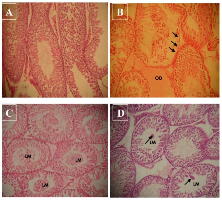
Figure 5.
Representative photomicrograph of the testicular sections showing the morphological changes induced by haloxyfop-p-methyl ester in the testes of rats (hematoxylin and eosin stain, 200×); control group (A) and treated groups at the doses of 6.75 mg/kg bw (B), 13.50 mg/kg bw (C) and 27.00 mg/kg bw (D). (A) shows normal testicular structure; in (B), there are severe interstitial edemas (OD) and some sections of the seminiferous tubule have necrotic and eroded germinal epithelium; in (C), most sections of the seminiferous tubules have immature cells in the lumen (LM); and in (D), there are few cellular clumps in the lumen of some of the seminiferous tubules.

