. 2017 Aug 21;6:e26653. doi: 10.7554/eLife.26653

Figure 5. Connectivity fingerprints of the subdivisions of Substantia Nigra.

The connectivity fingerprints of SN subdivisions were calculated based on a whole-brain atlas (Fan et al., 2016). The relative connectivity strength between each target (i.e. parcels in the brain atlas) and each SN subdivision is plotted. An organizational topography of SN projections emerges with vSN mostly connected to prefrontal cortex, lateral SNc to sensorimotor cortex, and medial SNc to limbic regions. The naming convention is based on Fan et al. (2016). The atlas is available at http://atlas.brainnetome.org/.

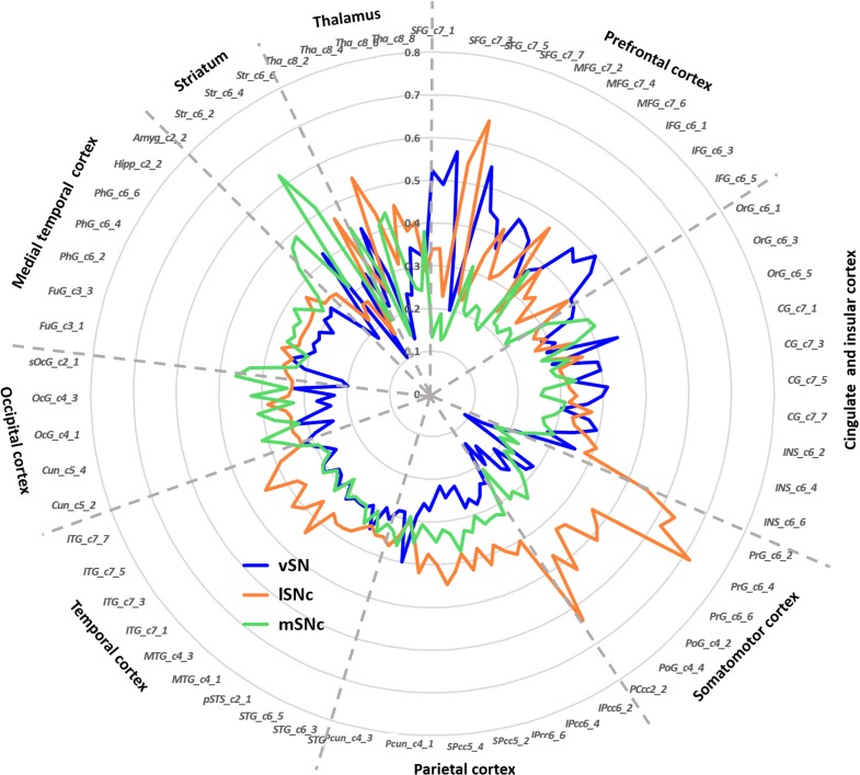
Figure 5.

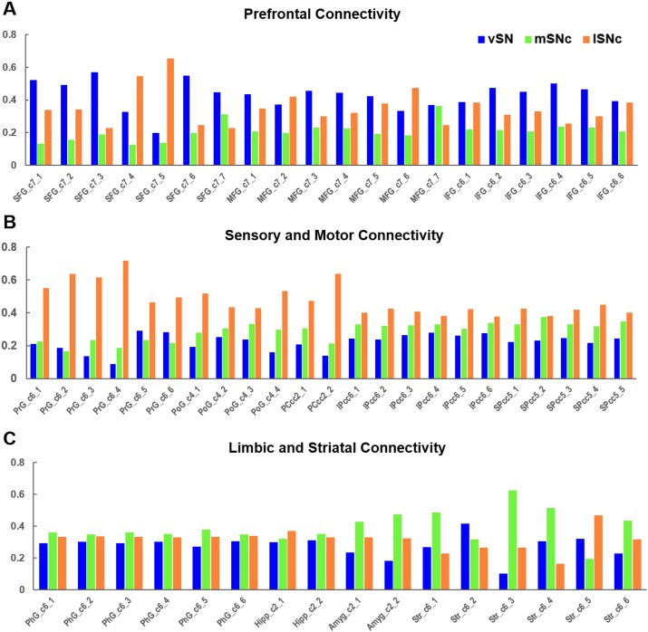
Figure 5—figure supplement 1. Connectivity profiles of SN subdivisions in prefrontal, motor and limbic systems.

Figure 5—figure supplement 1.

Organizational topography of SN projections showing that vSN preferentially connects with prefrontal cortex (A), lateral SNc with sensorimotor cortex (B), and medial SNc with limbic regions (C). Regions derived from the whole-brain atlas of (Fan et al., 2016).
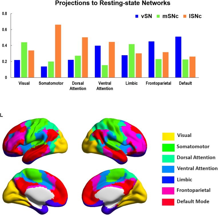
Figure 5—figure supplement 2. Connectivity profiles of SN subdivisions to seven resting-state functional networks.

Figure 5—figure supplement 2.

Organizational pattern of SN connections with seven canonical resting-sate networks (Yeo et al., 2011). A limbic-cognitive-motor topography was shown, where lSNc preferentially connects to somatomotor cortex and dorsal attention network; mSNc dominantly connects to limbic and visual cortex; vSN mainly connects with frontoparietal and default-mode networks. Bottom panel shows the cortical mapping of the seven resting-state networks by using BrainNetViewer (https://www.nitrc.org/projects/bnv).
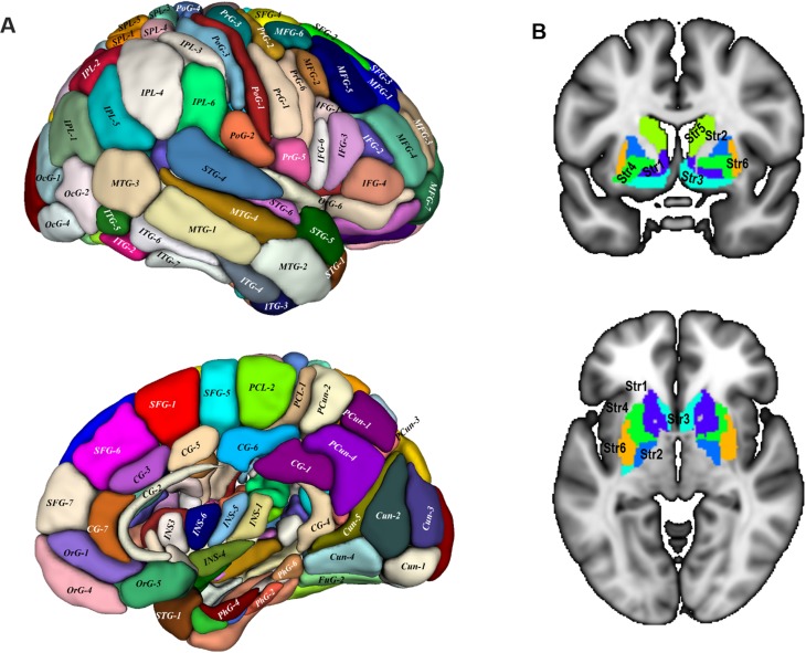
Figure 5—figure supplement 3. Target regions of the connectivity fingerprints.

Figure 5—figure supplement 3.

We used a whole-brain atlas (Fan et al., 2016) as the target areas to calculate the connectivity fingerprint of SN subdivisions. The surface view of the atlas (A) was adapted from (Fan et al., 2016) with permission from the authors. The slice views (B) depict the basal ganglia regions from the atlas. These are: Str1: ventral caudate, Str2: globus pallidus, Str3: nucleus accumbens; Str4: ventromedial putamen, Str5: dorsal caudate nucleus, Str6: dorsolateral putamen. This atlas is available at http://atlas.brainnetome.org/.