. 2017 Aug 21;6:e26653. doi: 10.7554/eLife.26653

Figure 6. Brain activity in response to rewarding and aversive outcomes in the fMRI gambling task.

Among SN subdivisions, only medial SNc showed a significant difference in response to reward and punishment (p<0.001). The ventral striatum (VS) and ventromedial prefrontal cortex (vmPFC) also responded differently to reward and punishment, with greater BOLD activity to rewarding than aversive stimuli (p<0.001). Meanwhile, anterior insula and dorsal anterior cingulate cortex (dACC) showed no difference in response to reward and punishment.

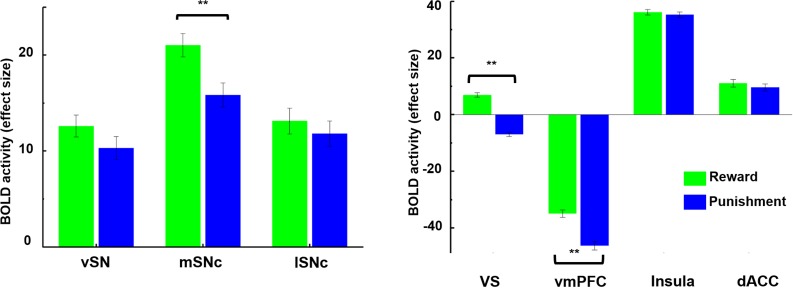
Figure 6.

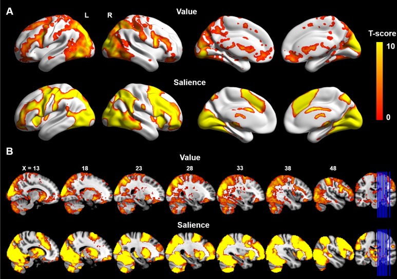
Figure 6—figure supplement 1. Whole-brain analysis of BOLD response to value-coding and salience-coding.

Figure 6—figure supplement 1.

One sample t-test was applied in FSL using the randomise function. Significant BOLD activity encoding value signals (top row; difference in response to reward and punishment) was detected in ventral medial prefrontal cortex and ventral striatum. Meanwhile, BOLD response to salience (second row; averaged response to reward and punishment) was found in anterior insula, dorsal ACC and dorsal striatum. A Threshold-Free Cluster Enhancement (TFCE) method (Smith and Nichols, 2009) was used to correct for multiple comparisons (FWE corrected p=0.01). A) Surface views generated using BrainNetViewer (https://www.nitrc.org/projects/bnv). (B) Multi-slice views generated using MRIcron (http://people.cas.sc.edu/rorden/mricron/index.html) on the ICBM152 brain template.
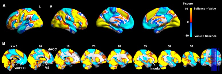
Figure 6—figure Supplement 2. Contrast between value- and salience-related BOLD responses.

Figure 6—figure Supplement 2.

Paired t-test was used to directly compare the contrasts of value and salience, correcting for multiple comparisons using the Threshold-Free Cluster Enhancement (TFCE) method (Smith and Nichols, 2009). Greater BOLD response to value (colored in blue) was observed in vmPFC, precuneus, hippocampus and ventral striatum. Greater BOLD response to salience (colored in yellow) was found in dorsal medial and lateral prefrontal, dorsal cingulate cortex, anterior insula and dorsal striatum.
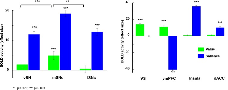
Figure 6—figure supplement 3. Brain activity in response to value and salience.

Figure 6—figure supplement 3.

Among the subdivisions of SN, only medial SNc significantly responded to value (gains > losses), and this was significantly greater than the other two subdivisions (p<0.001). However, all three subregions showed a significant response to salience (gains + losses) (p<0.0001). Ventral striatum and vmPFC both significantly responded to value (p<0.0001), but only vmPFC was deactivated by salience. Meanwhile, anterior insula and dorsal anterior cingulate cortex only significantly responded to salience. Overall, only mSNc, VS and vmPFC showed significant BOLD response to value, while all regions except VS responded to salience.