Fig. 4.

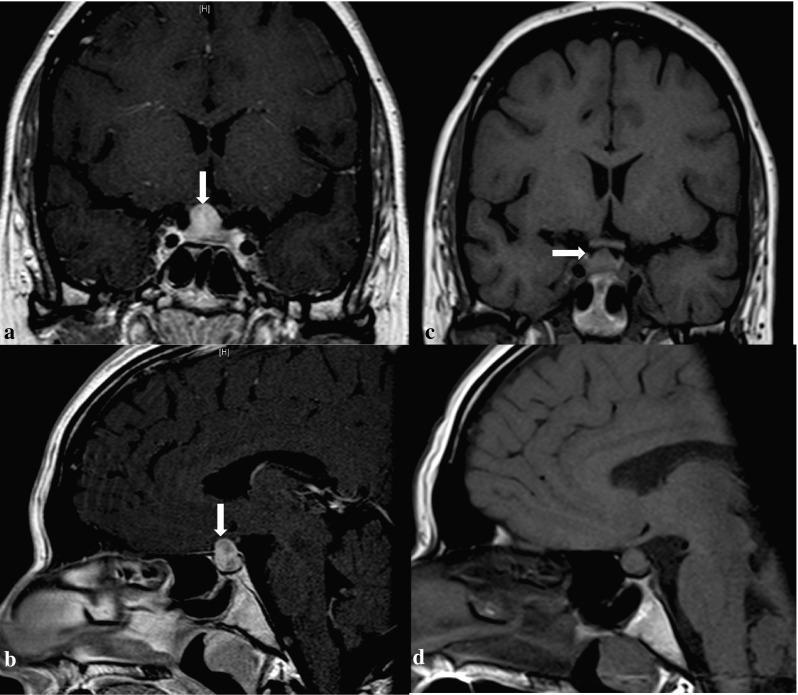
T1-weighted MRI (coronal and sagittal view) of a of 32-year-old woman (Case 3) with GPA associated PD showing a sellar mass with homogenous enhancement before (a and b) and after (c and d) treatment

T1-weighted MRI (coronal and sagittal view) of a of 32-year-old woman (Case 3) with GPA associated PD showing a sellar mass with homogenous enhancement before (a and b) and after (c and d) treatment