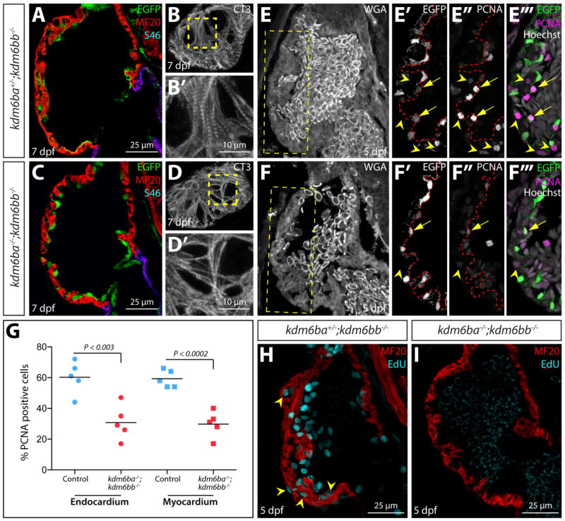
Figure 5. Kdm6b proteins enable coordinated endocardial and myocardial proliferation associated with early stage ventricle maturation.
(A–C) Confocal immunofluorescence images of sagittal heart sections from 5 dpf kdm6b- and kdm6a/kdm6b-deficient Tg(kdrl:EGFP) larvae stained with anti-GFP (green, endocardium), anti-myosin heavy chain (MF20, red, myocardium), and anti-S46 (magenta, atrial muscle) antibodies. (B, D) Cardiac troponin (CT3) antibody staining of superficial sections through the heart of 5 dpf kdm6bb−/− and kdm6ba−/−; kdm6bb−/− larvae. B′ and D′ are zoomed images of the dashed yellow box regions. (E–F) Confocal imaged sagittal sections of 5 dpf control and combined kdm6b-deficient Tg(kdrl:EGFP) embryos stained with wheat germ agglutinin (WGA, grey) as well as anti-GFP (green in overlay panels, endocardium) and anti-PCNA (magenta in overlay panels) antibodies. Yellow boxes in E and F indicate zoomed regions shown in E′–F‴. Arrows and arrowheads mark PCNA-positive endocardial and myocardial cells, respectively. (G) Scatterplot graphs showing the number of PCNA positive endocardial and myocardial cells within the outer curvature ventricular wall scored from matched sagittal heart sections of 5 dpf control and kdm6ba−/−; kdm6bb−/− larvae. Each point represents a distinct fish. P-values are from two-tailed Student’s t-tests. (H, I) Confocal heart images of sagittal sectioned 5 dpf control and kdm6a/kdm6b-deficient larvae stained for EdU incorporation (cyan, proliferating cells) and with anti-myosin heavy chain antibody (red, MF20, cardiomyocytes).

