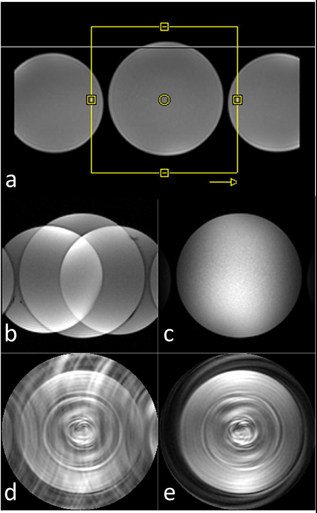
Figure 4.

OVS performance in phantoms (T1 = 100 ms) using Cartesian and spiral pulse sequences. (a) phantom setup along with a yellow-box representing the imaging FOV and an arrow indicating the phase encoding direction for the Cartesian pulse sequence. Cartesian reduced FOV images are shown without (b) and with (c) OVS preparation. Highly under-sampled spiral reduced FOV images are shown without (d) and with (e) OVS preparation.
