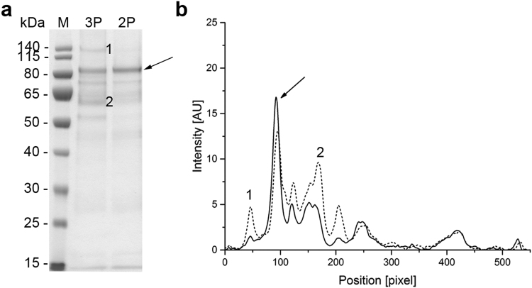
Figure 5.
The purity of VAMAX1 in the fermentation supernatant. (a) Proteins in 15 µL supernatant taken at the end of the three-phase (3 P) and two-phase (2 P) fermentation strategies were separated by LDS-PAGE and visualized with SimplyBlue SafeStain. (b) Densitometric analysis of the separated proteins using AIDA Image Analyzer. Arrows represent the target protein VAMAX1. Numbered bands/peaks represent impurities that were cut from the gel for analysis by mass spectrometry.

