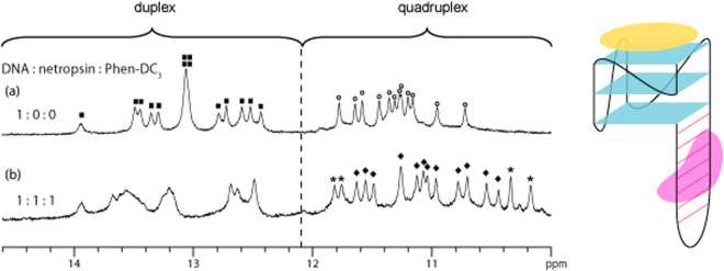
Figure 3.
1D imino proton NMR spectrum of (a) free QDH1 and (b) QDH1 bound with equimolar ratio of netropsin and Phen-DC3. Unbound and bound G-quadruplex imino proton peaks are labelled with open circles and filled diamonds, respectively, whereas duplex imino proton peaks of free QDH1 are labelled with filled squares. Peaks labelled with asterisks originate from the ligand Phen-DC3. Schematic structure of simultaneous netropsin (in pink) and Phen-DC3 (in yellow) binding to QDH1 is shown on the right.

