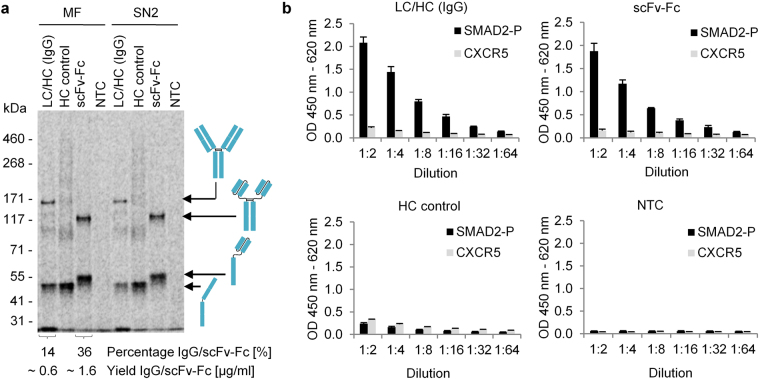
Figure 2.
Functional analysis of cell-free synthesized antibody constructs by ELISA. (a) Autoradiograph derived from SDS-PAGE gel showing cell-free synthesized target proteins in the microsomal fraction (MF) and the supernatant fraction (SN2) after detergent-based release of target proteins from the lumen of microsomal vesicles. (b) ELISA analysis showing the specific binding of LC/HC (IgG) and scFv-Fc to its antigen SMAD2-P, but not to CXCR5 (non-related antigen). Samples analyzed by ELISA were treated likewise and tested in parallel in triplicate analysis. Starting concentrations in dilution 1:2 of LC/HC, scFv-Fc and HC control were 0.2 µg/mL each (total protein yield according to quantification via incorporation of 14C-leucine). Standard deviations were calculated from triplicate analysis. LC: antibody light chain; HC: antibody heavy chain; scFv-Fc: single-chain variable fragment Fc fusion; NTC: No template control.

