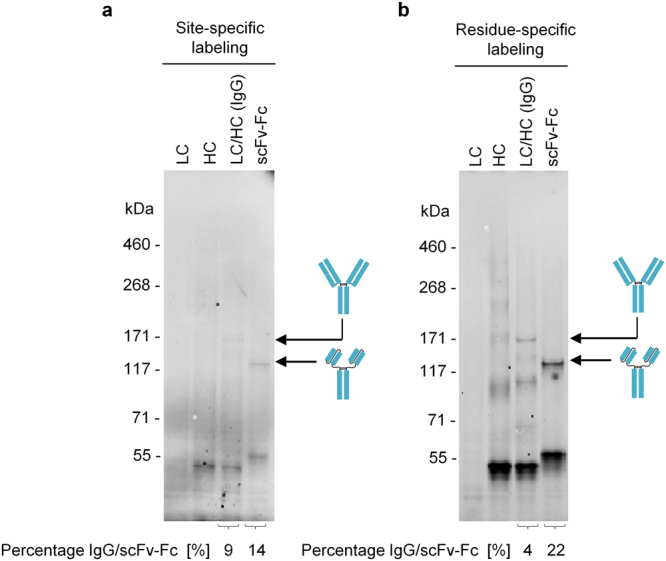
Figure 4.

In-gel-fluorescence of cell-free produced IgG and scFv-Fc labeled with Bodipy-TMR-lysine. Labeling was achieved by supplementing the batch-based cell-free reaction either with BODIPY-TMR-lysine-tRNACUA (site-specific labeling) or BODIPY-TMR-lysine-tRNAGAA (residue-specific labeling). (a) SDS-PAGE gel showing fluorescent protein bands of IgG and scFv-Fc in the microsomal fraction, site-specifically labeled at the position of the amber stop codon TAG located at the 3′-end of the open reading frame encoding HC as well as scFv-Fc template. (b) SDS-PAGE gel showing fluorescent protein bands of IgG and scFv-Fc (microsomal fraction) labeled by the incorporation of the fluorescent amino acid Bodipy-TMR-lysine at multiple sites in the protein (residue-specific labeling). LC: antibody light chain; HC: antibody heavy chain; scFv-Fc: single-chain variable fragment Fc fusion.
