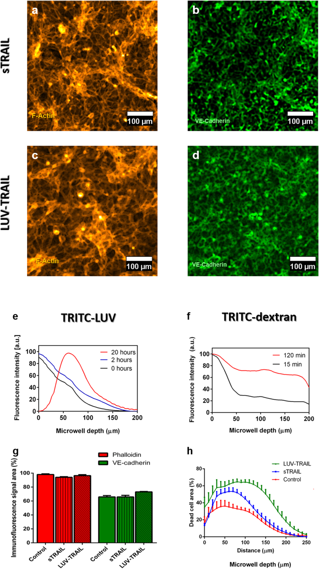
Figure 6.
Drug testing assay using TRAIL in both its soluble form and anchored to a LUV in the co-culture established models. Endothelium immunofluorescence after 24 h of exposure to 0.33 ng/ml sTRAIL (a,b) and 0.33 ng/ml LUV-TRAIL (c,d) for Phalloidin-TRITC (yellow) and VE-Cadherin (green), respectively. (e,f) Diffusion/penetration assays using TRITC-labelled LUV (4 mg/ml) and TRITC-Dextran (70 kDa, 30 µM) used as models for the LUV-TRAIL and s-TRAIL, respectively, across an endothelium seeded on a collagen gel. (g) Quantification of the endothelium integrity using the Phalloidin (red) and the VE-cadherin (green) signal area for control conditions (no TRAIL exposure) and samples treated with both sTRAIL and LUV-TRAIL (p-value > 0.99 as calculated through Kruskal-Wallis’ Test). (h) Quantification of MDA-MB-231 tumour cell death in 3D along the device microwell depth in control conditions, and with sTRAIL and LUV-TRAIL treatment in the established microfluidic co-culture model. Here, 5 million MDA-MB-231 cells/ml were seeded in the hydrogel matrix. Graphs show average ± SEM. N = 7.

