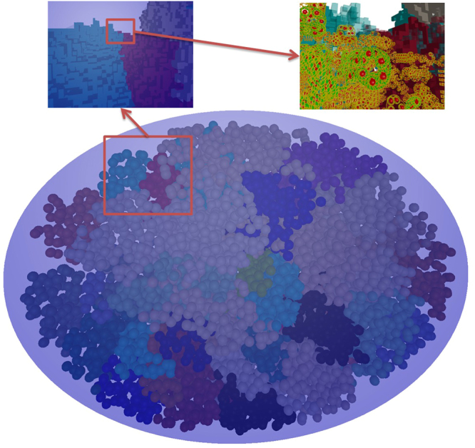
Figure 5.

A relaxed and filled fibroblast cell nucleus.The three views presented use three different levels of zoom. The main view (bottom) shows the whole nucleus from a distant point from which only the domains are visible. On the top left, the view is that of a zoom in on the red boxed area of the main view. At this level, it is possible to see the voxels as cubes. On the top right, the view comes from another zoom in on the red boxed area of the top left view. In this view, the detail of the DNA within the voxels is at a molecular level.
