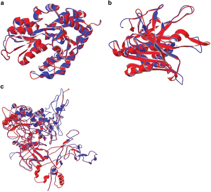
Figure 3.
Structure predictions for internal transcription sites versus PDB structures. (a) Protein structure threading for the cyanate ABC transporter (PDB. c2i4cA). There are no major structural differences in the protein threads. Blue and red are the overlapping structure representing the full length transcript (blue) and the corresponding predicted protein from the internal start site in MED4 (red). (b) As in A, but for the Ferredoxin-NADP reductase protein (PDB C1jb9A). (c) Protein structure threading for RNA polymerase (PDB C3lu0C).

