Figure 1.
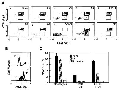
The L4 peptide induces positive selection of N15tg thymocytes. FTOC was performed by using N15tg/RAG-2−/−/β2m−/− (H-2b) thymic lobes in media containing 5 μg/ml human β2m with or without 10 μM of the indicated peptides. After 7 days, thymocytes were released from the lobes by pressing through a steel mesh, counted, and triple-stained with PE-conjugated anti-CD4, Red613-conjugated anti-CD8, and mAb R53 (anti-N15 β chain clonotype) plus FITC-conjugated anti-rat IgG (19). (A) The CD8 versus CD4 staining profile of total thymocytes is shown. In this representative experiment the yield of CD8+ SP cells after L4 incubation was 13.5 × 10⁴ cells compared with 0.3–2.2 × 10⁴ cells in FTOC incubated in the absence of exogenous peptides or in the presence of the other indicated peptides. The total thymocyte number recovered from VSV8-exposed FTOC was significantly lower (105 cells per lobe) than from FTOC incubated with any of the other peptides (4–6 × 10⁵ cells per lobe). (B) The histograms of the N15 TCRβ chain expression on DP and CD8+ SP thymocytes derived from FTOC incubated with K1 or L4 peptides are shown. Note that the K1 histogram represents data similar to that obtained with the other peptides (except VSV8). The CD8+ SP thymocytes that mature on L4 express a higher level of the TCR than the DP thymocytes harvested from the same lobe. (C) Thymocytes selected on L4 are functionally responsive to VSV8. Thymocytes from the organ cultures described above [cultured with (+) or without (−) L4] or fresh splenocytes from N15tg/RAG-2−/− mouse were assayed for their proliferative response to irradiated EL-4 cells, in the present of rIL-2 and 10 nM VSV8 or 10 μM L4 or no peptide. After 48 h, each well was pulsed for 18 h with [3H]thymidine, harvested on filter discs, and counted. The proliferative responses for the peptides are shown. Results are mean values of triplicate samples with SD noted.

