FIG 3.

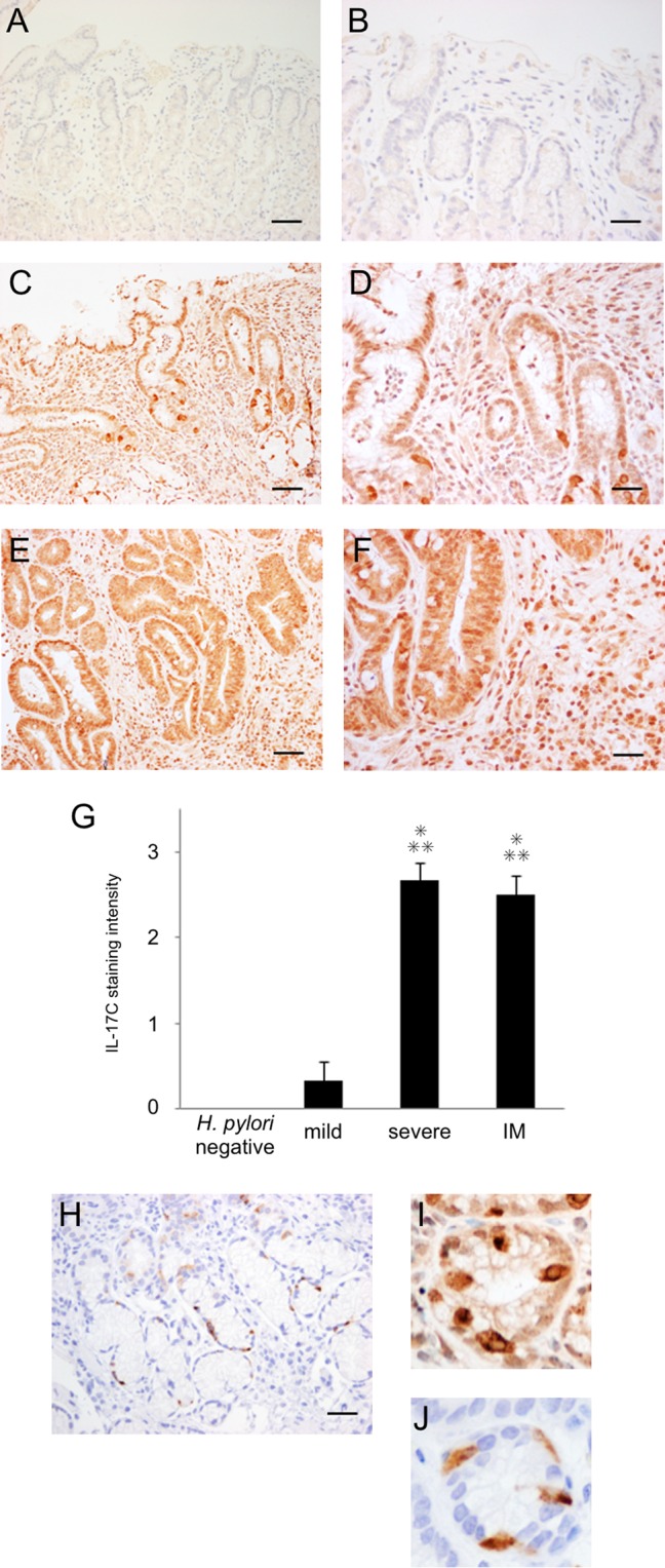
Immunohistochemistry of IL-17C (A to F and I) and chromogranin A (H and J) in the antral gastric mucosa. (A and B) Subject without H. pylori infection. (C, D, and H to J) Subject with H. pylori infection and severe gastritis. (E and F) Subjects with H. pylori infection and intestinal metaplasia. (G) Semiquantitative analysis of IL-17C. IL-17C staining was observed mainly in the epithelial cells of H. pylori-infected mucosa. Semiquantitative analysis of IL-17C in the gastric epithelial cells of H. pylori-negative and -positive subjects (no infection, n = 6; mild gastritis, n = 6; severe gastritis, n = 6; intestinal metaplasia [IM], n = 6) used the following scores: 0, no staining; 1, weak staining; 2, moderate staining; 3, strong staining (means ± standard errors [SE]; *, P < 0.001 compared with H. pylori-negative samples; **, P < 0.001 compared with mild gastritis). (I and J) Certain single cells in glandular epithelial cells were strongly stained, and these patterns resemble the staining of chromogranin A-positive enteroendocrine cells. Magnification, ×100 (scale bar, 250 μm) (A, C, E, and H) or ×400 (scale bar, 100 μm) (B, D, F, I, and J).
