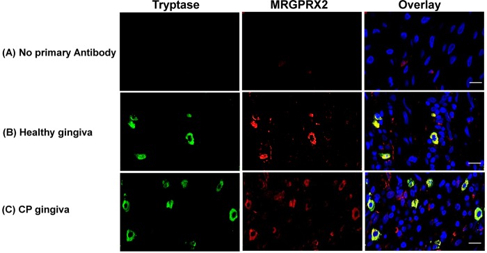
FIG 2.
Analysis of tryptase- and MRGPRX2-positive mast cells in periodontal tissue. Representative photomicrographs (n = 5 donors) of double immunofluorescence staining of gingival tissue are shown. Original magnification, ×60. (A) Control. (B and C) A healthy gingiva (B) and the gingiva of a patient with CP (C) were stained with anti-tryptase (green) and anti-MRGPRX2 (red) antibodies. Overlays of double-staining samples are shown. Bars = 50 μm.

