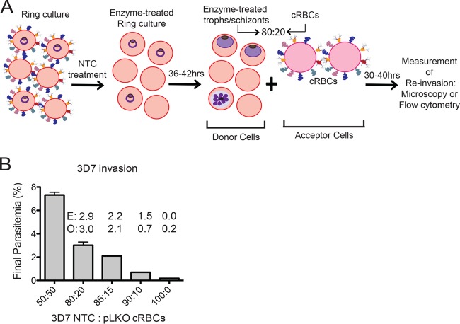
FIG 4.
Schematic of invasion assay design. (A) A ring-stage parasite culture is treated with neuraminidase, trypsin, and chymotrypsin (NTC), returned to culture conditions, and allowed to mature to the late trophozoite (troph) or schizont stage. The treated culture (donor cells) is mixed in an 80:20 ratio with either knockdown (KD) or control pLKO cRBCs (acceptor cells). Initial parasitemia and/or final parasitemia after one round of invasion are determined either by microscopy from counts of 500 to 2,000 erythrocytes or by flow cytometry of SYBR green I-stained cells. Invasion assays are set up at 0.5% hematocrit. All acceptor cells are counted by hemocytometer prior to assay setup. (B) Invasion of P. falciparum 3D7 treated with neuraminidase, trypsin, and chymotrypsin into pLKO control cRBCs with various ratio of enzyme-treated donor cells to pLKO acceptor cells. A 100:0 ratio indicates treated 3D7 donor cells and no pLKO cRBCs. Initial parasitemia was ∼2%. Final parasitemia was determined by flow cytometry of SYBR green I-stained cells. The assay was performed once in duplicate. Bars represent the mean ± the range. The 80:20 ratio was selected for subsequent invasion assays. E, expected parasitemia based on invasion into a 50:50 mixture. O, observed parasitemia.

