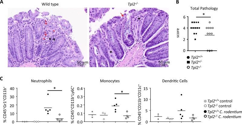
FIG 6.
Tpl2−/− mice have reduced neutrophil recruitment to the colon during the adaptive immune response. Wild-type and Tpl2−/− mice were gavaged with 2 × 10⁹ CFU Citrobacter rodentium (ICC180). Mice were euthanized at 8 or 11 dpi. (A) Representative images are given. The inflammatory cells in the colons at 11 dpi are mostly histiocytes (red dashed arrows) and lymphocytes (black solid arrows), with some neutrophils (red arrowheads) being seen. Asterisks denote areas where the intestinal glands are missing. The solid red arrow points to a colonic gland that is markedly dilated due to the accumulation of cellular debris and large numbers of bacteria in the lumen of the colonic gland. Magnifications, ×200. (B and C) Colons were isolated and scored for pathology (B), and the proportion of myeloid cells in the epithelial layer was quantified by gating on CD45+ events (C). Lines represent means (n ≥ 3 mice). (B) Data from three independent experiments were pooled. (C) Data are representative of those from one (monocytes and dendritic cells) or four (neutrophils) independent experiments. P values were determined by unpaired Student's t test (B) and one-way ANOVA (C). *, P < 0.05.

