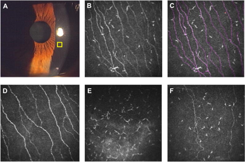
Figure 2. Laser in vivo confocal microscopy (IVCM) for the assessment of corneal nerves and dendritic cells.

Slit lamp microscopy is the gold standard for anterior segment examination to assess corneal infiltration, edema, corneal epithelial defects, and cells/flare in the anterior chamber (A). However, slit-lamp examination does not allow assessment of corneal nerves or dendritiform immune cells (DCs). Using laser IVCM, high-resolution images of the corneal cellular structures can be obtained in an area of 400 × 400 μm (yellow square). (B) IVCM image of a cornea with allergic conjunctivitis. (C) Using ImageJ/Neuron J, corneal nerve/DC density can be quantified. Corneal nerves are traced semi-automatically with Neuron J software (red lines). (D) IVCM image of a normal cornea. (E) IVCM image of an eye with microbial keratitis. Note the increased density of DCs, while the corneal nerve branches could not be detected. (F) IVCM image of the contralateral eye of a patient with unilateral microbial keratitis. Nerve density is decreased and DC density is increased compared with normal cornea.
