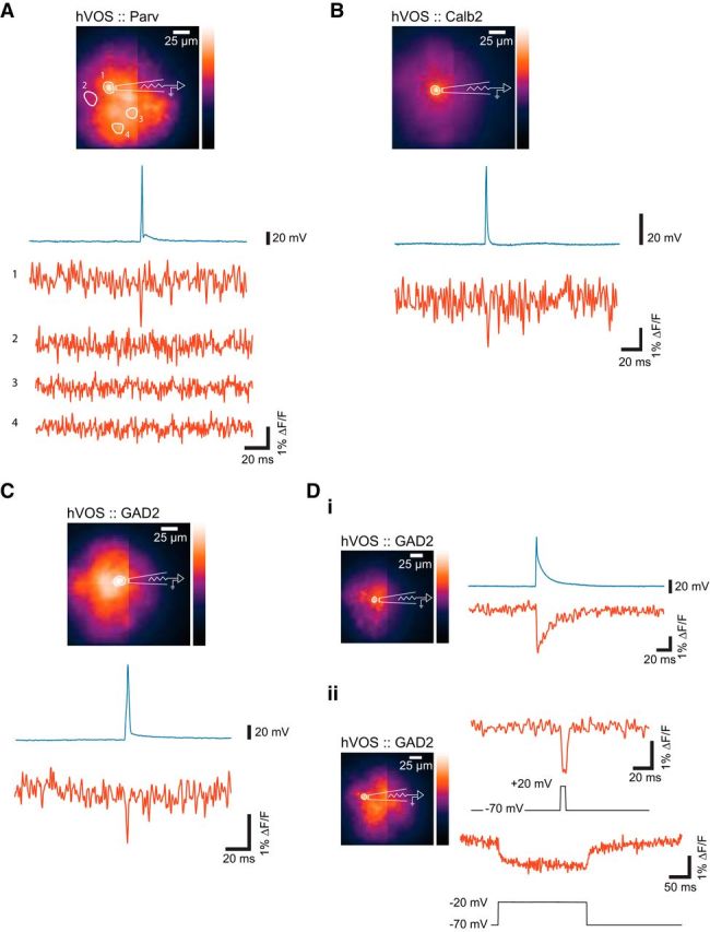
Figure 3.

Simultaneous imaging and electrical recording, with resting light images, whole-cell patch-clamp recordings (above), and hVOS responses (below). Action potentials were evoked by current injection under current clamp. A, Action potential and hVOS signal from a neuron in cortical layer 2/3 of a slice from an hVOS::Parv mouse. No fluorescence changes were seen in 3 other nearby cells numbered 2–4. B, Action potential and hVOS signal from a neuron in cortical layer 5 from an hVOS::Calb2 mouse. C, Action potential and hVOS signal from a layer 2/3 cortical neuron from an hVOS::GAD2 mouse. Di, hVOS and voltage responses to current injection in a GAD2 glial cell in layer 5 of the somatosensory cortex of an hVOS::GAD2 mouse. The cell was held at −30 mV. Dii, Glial cell hVOS responses to voltage steps (under voltage clamp) from −70 to 20 mV (5 ms, top) and −70 to −20 mV (200 ms, bottom traces) in layer 5 of the somatosensory cortex of an hVOS::GAD2 mouse. A–C, All hVOS recordings of neuronal responses are single trials. Glial responses are 5 trial averages, due to the significantly smaller signal amplitudes.
