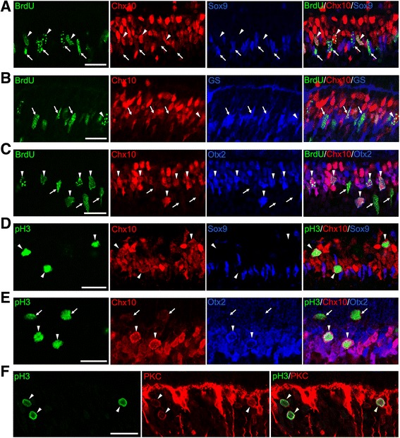
Fig. 4.

Ectopic cell cycle reentry of bipolar cells and Müller glia in the p27 knockout retinas at P9. a Triple immunofluorescence for BrdU, Chx10, and Sox9. Arrows indicate BrdU+ cells stained weakly for Chx10 and intensely for Sox9 (Müller glia) while arrowheads denote BrdU+ cells which are intensely Chx10+ and Sox9- (bipolar cells). b Triple immunofluorescence for BrdU, Chx10, and glutamine synthetase (GS). Arrows indicate BrdU+, weakly Chx10+ and GS+ cells (Müller glia). Arrowhead shows BrdU+, intensely Chx10+, and GS- cell (bipolar cell). c Triple immunofluorescence for BrdU, Chx10, and Otx2. Some BrdU+ cells are weakly Chx10+ and Otx2- (arrows, Müller glia) while others are intensely positive for both Chx10 and Otx2 (arrowheads, bipolar cells). d Triple immunofluorescence for phospho-histone H3 (pH3), Chx10, and Sox9. Arrowheads indicate ectopic M-phase cells strongly positive for Chx10, but negative for Sox9 (bipolar cells). e Triple immunofluorescence for pH3, Chx10, and Otx2. Arrows denote pH3+ cells stained weakly for Chx10 and negative for Otx2 (Müller glia). Arrowheads indicate pH3+/Chx10+/Otx2+ cells (bipolar cells). f Double immunofluorescence for pH3 and PKCα (PKC) showing colocalization (arrowheads, bipolar cells). Scale bar = 20 μm
