Fig. 8.

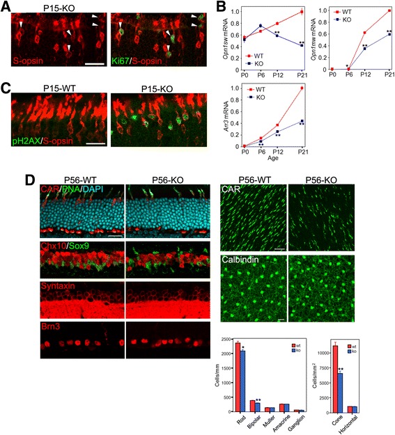
Impaired differentiation and survival of cones in the p27 knockout (KO) retinas. a Double immunofluorescence for Ki67 and S-opsin in the KO retina at P15. Note relatively weak S-opsin labeling in the Ki67+ cones (arrowheads). b Quantitative RT-PCR analyses of cone gene expression in the WT and KO retinas during postnatal development. The transcript levels are expressed relative to WT at P21 after normalized to Gapdh levels. Each value represents the mean ± SEM (n = 3 per stage and genotype). *P < 0.05, **P < 0.01, Student’s t test. c Double immunofluorescence for phospho-H2AX (pH2AX) and S-opsin in the WT and KO retinas at P15. Note pH2AX+ cones in the KO retina. d Quantification of retinal cell types in the WT and p27KO retinas at P56. Retinal sections or whole mounts were immunolabeled for cell-specific markers. Note a significant reduction in cone number in the KO retina. The number of rods and bipolar cells are also mildly reduced. Bars represent the mean ± SEM (n= 3 per genotype). *p < 0.05, **p < 0.01, Student’s t test. Scale bars are 20 μm
